Anatomy Muscles Pelvis ~ The Pelvic Floor Structure Function Muscles Teachmeanatomy. The pubococcygeus (pc) muscle is the muscle that runs the show in pelvic floor health. These ligaments reinforce and stabilize the hip joint (6). This mri male pelvis axial cross sectional anatomy tool is absolutely free to use. The gluteal muscles are a group of three muscles named the gluteus maximus, the gluteus medius, and the gluteus minimus. Anatomy of pelvic and acetabular muscles.
Large ligaments, tendons, and muscles around the hip joint hold the bones (ball and socket) in place and keep it from dislocating. The pelvic floor muscles provide foundational support for the intestines and bladder. Use the mouse scroll wheel to move the images up and down alternatively use the tiny arrows (>>) on both side of the image to move the images.>>) on both side of the image to move the images. The thigh bone or femur and the pelvis join to form the hip joint. Several muscles around the pelvis take part in movements of the thigh.
Your Pelvic Floor Muscles Why You Should Care Our Fit Family Life from images.squarespace-cdn.com The iliofemoral, pubofemoral, and ischiofemoral ligaments represent the thickenings of the joint capsule. The hip joint is an intricate structure including hip bones, hip articular cartilage, muscles, ligaments and tendons, and synovial fluid. The tensor fasciae latae (tfl) is a small muscle on the outside of the hip. The levator ani muscles consist of three. See more ideas about pelvis anatomy, anatomy, pelvis. It is usually divided into two separate anatomic regions: The gluteal muscles are a group of three muscles named the gluteus maximus, the gluteus medius, and the gluteus minimus. The pelvic girdle, also known as the hip bone, is composed of three fused bones:
The term pelvis is used to identify the area between the abdomen and the lower extremities.
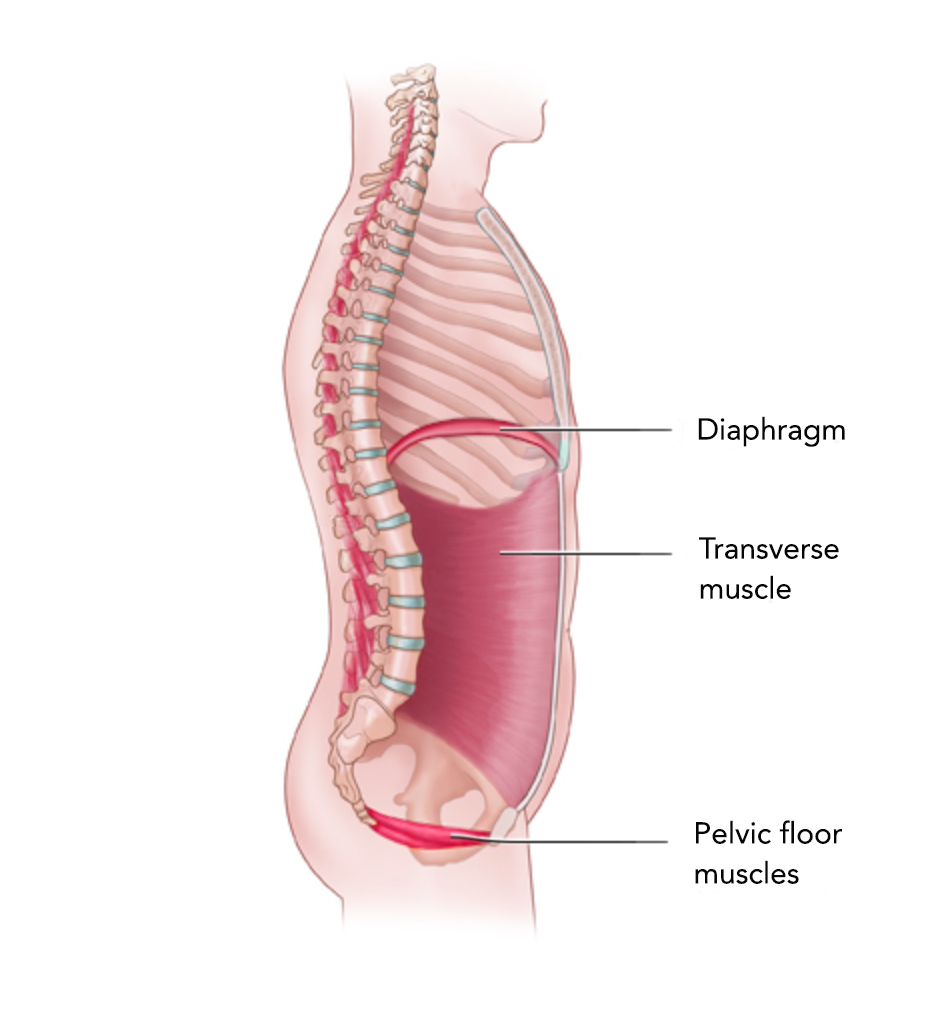
Muscles an important group of muscles in the pelvis is the pelvic floor. Each of the hip muscles will have a main function, to produce a specific movement. These muscles, including the gluteus maximus and the hamstrings, extend the thigh at the hip in support of the body's weight and propulsion. It is usually divided into two separate anatomic regions: The iliacus muscle interacts with the bundles of the abdominal muscle between your lowest rib and the top of your pelvis (quadratus lumborum muscle). The pelvis also houses the reproductive organs, which have their own muscles. The pelvis consists of the sacrum, the coccyx, the ischium, the ilium, and the pubis. Several muscles around the pelvis take part in movements of the thigh. This mri male pelvis axial cross sectional anatomy tool is absolutely free to use. The muscles of the pelvic floor are collectively referred to as the levator ani and coccygeus muscles. The pubococcygeus (pc) muscle is the muscle that runs the show in pelvic floor health. The levator ani is a broad sheet of muscle. The rectus femoris is the most superficial of the quadriceps muscles alongside the vastus lateralis, vastus intermedius, and vastus medialis.
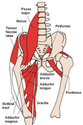
The adductor muscle group, also known as the groin muscles, is a group located on the medial side of the thigh. This mri male pelvis axial cross sectional anatomy tool is absolutely free to use. It is also referred to as a ball and socket joint and is surrounded by muscles, ligaments, and tendons. Muscles that attach from the pelvis to the trunk and cross the lumbosacral joint muscles that attach from the pelvis to the thigh/leg and cross the hip joint pelvic floor muscles that are located wholly within the pelvis Anatomy of pelvic and acetabular muscles.
Pelvis Wikipedia from upload.wikimedia.org They have several functions, including helping to support the pelvic organs. This mri male pelvis axial cross sectional anatomy tool is absolutely free to use. The pelvis also houses the reproductive organs, which have their own muscles. The levator ani is a broad sheet of muscle. These ligaments reinforce and stabilize the hip joint (6). These muscles have attachments to the pelvis as follows: Included in this group are the adductor longus, adductor brevis, adductor magnus, pectineus, and gracilis muscles. The pelvis consists of the sacrum, the coccyx, the ischium, the ilium, and the pubis.
These muscles move the thigh toward the body's midline.
The thigh bone or femur and the pelvis join to form the hip joint. The iliofemoral, pubofemoral, and ischiofemoral ligaments represent the thickenings of the joint capsule. Anatomy of pelvic and acetabular muscles. The muscles of the pelvis form its floor. The hip joint is an intricate structure including hip bones, hip articular cartilage, muscles, ligaments and tendons, and synovial fluid. The tensor fasciae latae (tfl) is a small muscle on the outside of the hip. Some of the major pelvic muscles are as follows. The pelvic girdle and pelvic spine. These four muscles conjoin to attach to the patella as the quadriceps tendon. There are many muscles that form the pelvic floor, including puborectalis, pubococcygeus, iliococcygeus and coccygeus. It can be divided into the greater pelvis and the lesser pelvis. The muscles of the pelvic floor are collectively referred to as the levator ani and coccygeus muscles. The levator ani muscles are the largest group of muscles in the pelvis.
The ilium, ischium and the pubic bone. The hip joint is made up of two bones: The hip joint is an intricate structure including hip bones, hip articular cartilage, muscles, ligaments and tendons, and synovial fluid. They form a large sheet of skeletal muscle that is thicker in some areas than in others. The sacrum, five fused vertebral bones, joins the pelvis between the crests of the ilium.
Muscles Of The Pelvis from www.learnmuscles.com Included in this group are the adductor longus, adductor brevis, adductor magnus, pectineus, and gracilis muscles. Below the sacrum is the coccyx, or tailbone, a section of fused bone that is the end of the vertebral. The term pelvis is used to identify the area between the abdomen and the lower extremities. There are many muscles that form the pelvic floor, including puborectalis, pubococcygeus, iliococcygeus and coccygeus. The levator ani muscles are the largest group of muscles in the pelvis. The bony pelvis consists of the two hip bones (also known as innominate or pelvic bones), the sacrum and the coccyx. It is usually divided into two separate anatomic regions: The muscles of the pelvis form its floor.
There are four articulations within the pelvis:
The hip joint is made up of two bones: Included in this group are the adductor longus, adductor brevis, adductor magnus, pectineus, and gracilis muscles. The pelvis and the femur (the thighbone). They form a large sheet of skeletal muscle that is thicker in some areas than in others. Muscles that attach from the pelvis to the trunk and cross the lumbosacral joint muscles that attach from the pelvis to the thigh/leg and cross the hip joint pelvic floor muscles that are located wholly within the pelvis The hip's essential muscles are the sartorius, rectus femoris, gluteus minimus and medius, iliopsoas, adductors, and hamstrings. The pubococcygeus (pc) muscle is the muscle that runs the show in pelvic floor health. The iliofemoral, pubofemoral, and ischiofemoral ligaments represent the thickenings of the joint capsule. The ball is the rounded end of the femur (also called the. The tensor fasciae latae (tfl) is a small muscle on the outside of the hip. The floor of the pelvis is made up of the muscles of the pelvis, which support its contents and maintain urinary and faecal continence. The pelvic girdle, also known as the hip bone, is composed of three fused bones: Each of the hip muscles will have a main function, to produce a specific movement.
Share :
Post a Comment
for "Anatomy Muscles Pelvis ~ The Pelvic Floor Structure Function Muscles Teachmeanatomy"


Post a Comment for "Anatomy Muscles Pelvis ~ The Pelvic Floor Structure Function Muscles Teachmeanatomy"